
Scope and Framing
This analysis evaluates the central thesis of Kim and Munster’s commentary (Kim & Munster, J Clin Oncol 2026) — that estrogen alone is not carcinogenic for breast cancer and may be protective, with progestogens being the “true culprit” — by examining whether the cited evidence supports their claims and identifying major contradictory evidence the paper omits. The paper makes several arguments that are partially supported by legitimate data but ultimately presents a selective reading of the evidence that omits critical counterarguments. Throughout, the paper relies almost exclusively on relative risk reductions (“22% reduction,” “40% reduction in mortality”) without providing absolute risk differences or baseline event rates, a framing that systematically overstates clinical effect sizes.
Part I: Evaluation of the Paper’s Key Arguments and Cited Evidence
- The WHI Estrogen-Only Trial: Accurately Cited but Incompletely Contextualized
The paper’s strongest evidence is the WHI conjugated equine estrogen (CEE)-alone trial, and the cited numbers are accurate: estrogen-only therapy reduced breast cancer incidence by 22% (HR 0.78; 95% CI 0.65–0.93) and breast cancer mortality by 40% (HR 0.60; 95% CI 0.37–0.97) at more than 20 years of follow-up (Chlebowski et al., JAMA 2020). These findings have been consistent across serial reports and are supported by a meta-analysis of 10 randomized trials (RR 0.77; 95% CI 0.65–0.91) (Chlebowski et al., Breast Cancer Res Treat 2024). The divergence between the CEE-alone arm and the CEE plus medroxyprogesterone acetate (MPA) arm (HR 1.28; 95% CI 1.13–1.45 for incidence) is genuinely striking and does support a role for progestins in driving the excess risk seen with combined therapy (Chlebowski et al., JAMA 2020).
The paper does not, however, convey the caveats that limit generalizability:
– The CEE-alone arm enrolled only hysterectomized women (n=10,739), approximately 40% of whom had undergone prior bilateral oophorectomy — itself associated with reduced breast cancer risk. This population differs fundamentally from typical menopausal hormone therapy users (Gompel & Simcock, Lancet Diabetes Endocrinol 2026).
– Mean age at enrollment was 63.6 years, with roughly half of participants 20 or more years past menopause (Chlebowski et al., JAMA 2020; Manson et al., JAMA 2024). Under the timing or gap-time hypothesis, initiating estrogen near the menopausal transition — when breast tissue has not undergone years of near-complete estrogen deprivation — represents a different biological setting from initiation 10 to 20 years post-menopause. Re-introduction after prolonged deprivation may induce apoptosis in adapted pre-neoplastic cells, whereas initiation near menopause supports epithelial survival (Jordan, Endocr Relat Cancer 2015).
– Approximately 85% of breast cancer cases occurred in women who were overweight or obese, in whom adipose-derived estrogen already provides substantial estrogenic stimulation, attenuating any incremental effect of exogenous estrogen (Collaborative Group on Hormonal Factors in Breast Cancer, Lancet 2019).
– At 21 years of cumulative follow-up the mortality reduction lost statistical significance (RR 0.67; 95% CI 0.42–1.05), raising questions about durability (Bofill Rodriguez et al., Cochrane Database Syst Rev 2025).
– CEE contains more than 200 constituents, including equine-specific estrogens with pro-apoptotic properties not shared by estradiol-based formulations (Gompel & Simcock, Lancet Diabetes Endocrinol 2026; Yue et al., Int J Cancer 2018).
– In absolute terms the effect was modest: 238 versus 296 breast cancer cases (CEE versus placebo), approximately 5 to 6 fewer cases per 10,000 person-years, and 30 versus 46 breast cancer deaths (Gompel & Simcock, Lancet Diabetes Endocrinol 2026; Manson et al., JAMA 2024).
The WHI finding is real but may reflect a specific biological context — estrogen re-introduction after prolonged deprivation in older, frequently obese, post-hysterectomy women — rather than evidence that estrogen is universally non-carcinogenic.
- Progestin-Only Versus Combined Contraceptives and Bioidentical Progesterone: Oversimplified
The paper states that women using progestin-only contraceptives show higher breast cancer risk than those using combined pills, presenting this as evidence that estrogen is not the carcinogenic component. The claim is partially supported but substantially oversimplified.
Fitzpatrick and colleagues concluded that risks were similar in magnitude across methods: combined oral OR 1.23, progestin-only oral OR 1.26, and progestin-releasing intrauterine devices OR 1.32 (Fitzpatrick et al., PLoS Med 2023). The largest study to date did find a significantly higher hazard for progestin-only (1.21) than combined preparations (1.12; Z-test P=0.006), but median duration of use was approximately 40% longer among progestin-only users, and risk varied more by progestin type — desogestrel exceeding levonorgestrel, exceeding drospirenone and depot medroxyprogesterone acetate — than by the combined versus progestin-only distinction (Hadizadeh et al., JAMA Oncol 2025). Channeling bias is a recognized confounder the paper does not acknowledge: progestin-only methods are preferentially prescribed to women with contraindications to estrogen, including obesity, smoking, and older age. A Swedish register study of 1.5 million women found absolute risk per 100,000 woman-years was lower among current users of combined contraception (10.9) than progestin-only users (29.8), with the authors explicitly attributing part of this to selective prescription to women with breast cancer risk factors (Niemeyer Hultstrand et al., Lancet Reg Health Eur 2022).
The paper further argues that bioidentical micronized progesterone carries lower risk than synthetic progestins. In the French E3N cohort of 78,353 postmenopausal women, short-term use (under 5 years) of micronized progesterone with estrogen was not significantly associated with increased risk (HR 1.13; 95% CI 0.99–1.29), whereas use beyond 5 years significantly elevated risk (HR 1.31; 95% CI 1.15–1.48) (Fournier et al., Breast Cancer Res Treat 2014). Micronized progesterone exhibits lower mitotic potency than synthetic progestins but is not endocrinologically inert over extended durations.
- Systemic Therapy Versus Local Vaginal Estrogen: Distinct Clinical Entities
The paper cites observational data showing that vaginal estrogen therapy for genitourinary symptoms in breast cancer survivors was not associated with increased breast cancer mortality (HR 0.77; 95% CI 0.63–0.94) (McVicker et al., JAMA Oncol 2024). Low-dose vaginal preparations produce minimal and transient systemic absorption, generally keeping serum estradiol within the postmenopausal reference range. Differentiating local from systemic therapy explains why vaginal estrogen can be considered for genitourinary symptoms without undermining evidence that systemic estrogen promotes ER-positive proliferation — and it means these data cannot be recruited, as the paper implicitly does, to support the safety of systemic estrogen.
- Tamoxifen’s Mechanism: A Plausible but Incomplete Reframing
The argument that tamoxifen acts partly by blocking estrogen-induced progesterone receptor (PR) expression is biologically plausible and consistent with the cited data showing greater benefit in ER+/PR+ than ER+/PR− tumors. The Italian prevention trial finding — risk reduction confined to women concurrently using estrogen-only therapy — is accurately cited.
The reframing nonetheless understates that tamoxifen’s principal mechanism is competitive antagonism at ERα, directly blocking estrogen-driven transcription and proliferation (Burstein, N Engl J Med 2020). The greater benefit in ER+/PR+ disease is more parsimoniously explained by PR expression serving as a marker of a functionally intact ER signaling pathway, and therefore of greater sensitivity to ER blockade, than by PR signaling being the primary target. The Italian trial enrolled only hysterectomized women, and the interaction with hormone therapy use has not been replicated in the larger NSABP P-1, IBIS-I, or Royal Marsden prevention trials.
- BRCA1 Carriers and Hormone Therapy: Accurately Cited but Conflates Tumor ER Status With Initiation Biology
Data showing no adverse effect of estrogen-only therapy after oophorectomy in BRCA1 carriers, with increased risk from estrogen-progestin therapy, are accurately represented (Regev-Sadeh et al., JAMA Netw Open 2026) and do support a progestin-driven mechanism in this population.
The paper’s use of ER-negative tumor mortality reductions after oophorectomy to argue that estrogen is inert contains a logical flaw. BRCA1-associated cancers are 70% to 85% triple-negative or basal-like by intrinsic biology, whereas BRCA2-associated cancers are approximately 75% to 80% ER-positive (Breast Cancer Association Consortium, JAMA Oncol 2022; Tung et al., J Clin Oncol 2020). The argument conflates the ER status of the resulting tumor with the hormonal milieu required for initiation. In BRCA1-deficient mouse models, mammary tumors are ERα-negative, yet bilateral ovariectomy abrogates tumor development and hormonal replacement experiments show tumor formation is promoted by estrogen rather than progesterone (van de Ven et al., J Pathol 2018). Estrogen metabolites cause DNA double-strand breaks in ER-negative BRCA1-mutant cells, and BRCA1 deficiency exacerbates this genotoxicity because BRCA1 is required to repair such lesions (Savage et al., Cancer Res 2014). ERα may also be expressed during early BRCA1-associated tumorigenesis and lost during progression (Li et al., Oncogene 2007; Jones et al., Oncogene 2008). The observation is therefore consistent with estrogen carcinogenesis operating through genotoxic metabolites and early ER-dependent initiation, not evidence against it.
- IVF and Ovarian Stimulation: Selective Interpretation and an Incorrect Endocrine Premise
The paper cites reduced breast cancer recurrence following ovarian stimulation as evidence that high estrogen is protective. Meta-analytic data do show no increased recurrence risk, and possibly a reduction, with fertility preservation (Arecco et al., Hum Reprod 2022). The inference nonetheless fails on two grounds.
First, the paper’s endocrine premise is incomplete. It characterizes ovarian stimulation as a high-estrogen state accompanied by impaired progesterone synthesis from defective corpus luteum formation. Luteal-phase deficiency after stimulation is precisely why standard assisted reproduction protocols administer substantial exogenous progesterone — vaginal, intramuscular, or oral — from oocyte retrieval through early gestation (Garg et al., Nat Rev Endocrinol 2024; Mohammed et al., Fertil Steril 2019). Patients undergoing stimulation are therefore not reliably progesterone-deplete; many are progesterone-replete by supplementation. The cohort cannot serve as a natural experiment isolating estrogen from progestogen exposure.
Second, even accepting a period of relative progesterone deficiency, the confounding of supraphysiologic estrogen with altered progesterone makes attribution to estrogen alone impossible. The population is additionally highly selected — younger, healthier, with favorable prognostic features and closer surveillance — introducing selection bias that meta-analysis of observational cohorts cannot remove (Arecco et al., Hum Reprod 2022).
- The Equivalence Fallacy: Estrogen-Induced Apoptosis in LTED Cells Versus Intact Epithelium
The paper invokes estrogen-induced apoptosis in long-term estrogen-deprived (LTED) breast cancer cell lines to argue that estrogen is broadly protective, extending the mechanism to the WHI finding, high-dose estrogen therapy, and IVF-associated estrogen surges. This conflates two biologically distinct contexts.
– In LTED and aromatase inhibitor-resistant ER-positive cancer cells, estrogen at physiologic concentrations triggers apoptosis through acquired adaptations: Fas receptor upregulation, AMPK activation (which estradiol inhibits in wild-type cells), and endoplasmic reticulum stress via PERK and the unfolded protein response (Song et al., J Natl Cancer Inst 2001; Ariazi et al., Proc Natl Acad Sci U S A 2011; Chen et al., Apoptosis 2015; Sengupta et al., Mol Cancer Res 2019). A minimum interval of roughly 5 years of deprivation appears necessary to select cell populations vulnerable to this effect (Jordan, Endocr Relat Cancer 2015).
– In normal, non-deprived mammary epithelium, estrogen acts as a mitogen and survival factor. Human breast tissue organ cultures show estradiol increases proliferation and decreases apoptosis, and normal breast epithelial cells upregulate anti-apoptotic Bcl-2 in response (Eigeliene et al., BMC Cancer 2006; Somaï et al., Int J Cancer 2003; Gompel et al., Steroids 2000).
Receptor subtype further undermines any uniform reading of estrogen action. ERα and ERβ exert substantially opposing effects in mammary tissue: ERβ inhibits proliferation and induces G2 cell-cycle arrest in breast cancer cells, and promotes anti-proliferative and pro-apoptotic programs in mammary epithelium (Paruthiyil et al., Cancer Res 2004; Helguero et al., Oncogene 2005). This does not rescue the paper’s thesis. ERβ’s growth-inhibitory effects are themselves repressed by MAPK and PI3K activation, and its role in breast cancer remains genuinely contested rather than reliably protective (Cotrim et al., Oncogene 2013; Shen et al., Pharmacol Ther 2023). The relevant point is that a claim about “estrogen” is underspecified: net tissue effect depends on ERα/ERβ ratio, ligand, and cellular context, and the paper’s protective signals arise in settings where that ratio and the prior deprivation state are not characterized.
The molecular prerequisites for estrogen-induced apoptosis — prolonged near-complete deprivation, malignant transformation, Fas expression, altered AMPK signaling — are absent in normal postmenopausal breast tissue, which retains measurable estrogen from peripheral aromatization (Gruber et al., N Engl J Med 2002). Extrapolating from LTED cancer biology to the claim that estrogen-only therapy lowers risk by inducing apoptosis in postmenopausal breast tissue is mechanistically unsupported. CEE additionally shows pro-apoptotic properties distinct from estradiol in animal models, so the WHI result may partly reflect CEE-specific pharmacology (Yue et al., Int J Cancer 2018; Santen & Yue, Climacteric 2019).
Part II: Major Contradictory Evidence Not Cited
- Aromatase Inhibitor Prevention Trials — The Most Critical Omission
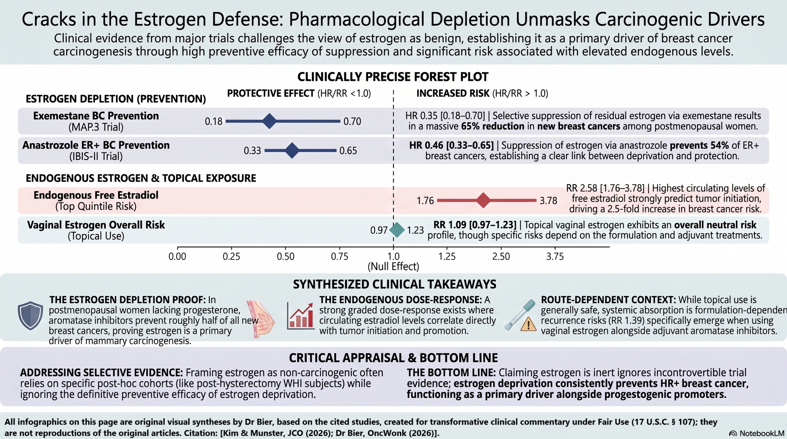
The paper does not cite or discuss aromatase inhibitor data at all, the single most important gap in its argument. Aromatase inhibitors act by reducing estrogen synthesis, suppressing residual estrogen by more than 90% in postmenopausal women (Burstein, N Engl J Med 2020). In postmenopausal women, where progesterone is negligible, they prevent approximately half of new ER-positive cancers:
– MAP.3: exemestane reduced invasive breast cancer by 65% versus placebo (HR 0.35; 95% CI 0.18–0.70) (Goss et al., N Engl J Med 2011).
– IBIS-II: anastrozole reduced all breast cancer by 49% at 131 months (HR 0.51; 95% CI 0.39–0.66), with benefit persisting after cessation (HR 0.64; 95% CI 0.45–0.91) (Cuzick et al., Lancet 2020).
– Cochrane network meta-analysis: aromatase inhibitors reduced overall breast cancer by 53% versus placebo (RR 0.47; 95% CI 0.35–0.63; high-certainty evidence) (Mocellin et al., Cochrane Database Syst Rev 2019).
If progestogens were the sole driver and estrogen inert, aromatase inhibitors should have no preventive effect in a population with negligible progesterone. A case-control analysis within IBIS-II further showed anastrozole’s benefit concentrated among women with higher baseline estradiol — a dose-response relationship linking endogenous estrogen directly to efficacy (Cuzick et al., Lancet Oncol 2024).
- Aromatase Inhibitors in Adjuvant Treatment
The EBCTCG patient-level meta-analysis of 31,920 women showed aromatase inhibitors reduced recurrence by approximately 30% and 10-year breast cancer mortality by approximately 15% relative to tamoxifen (EBCTCG, Lancet 2015). Extended therapy demonstrated a duration-dependent gradient, with longer estrogen suppression producing greater recurrence reduction (trend P<0.00001) (EBCTCG, Lancet 2025). A pharmacological dose-response relationship of this kind is a recognized indicator of causality.
- Ovarian Suppression Trials
SOFT and TEXT demonstrated stepwise benefit from progressively deeper estrogen suppression in premenopausal women: 15-year breast cancer-free interval was 72.1% with tamoxifen alone, 75.7% with tamoxifen plus ovarian function suppression, and 78.6% with exemestane plus suppression. Among women under 35 with HER2-negative disease, the 15-year overall survival difference reached 14.4 percentage points favoring maximal suppression (82.5% versus 68.1%) (Francis et al., Ann Oncol 2026). The 2026 EBCTCG meta-analysis of 15,075 women across 23 trials confirmed that ovarian suppression significantly reduced recurrence (RR 0.82; 95% CI 0.77–0.87), including when added to tamoxifen (EBCTCG, Lancet 2026).
- Endogenous Estrogen Levels and Breast Cancer Risk
The Endogenous Hormones and Breast Cancer Collaborative Group pooled analysis of 9 prospective studies found a graded dose-response, with women in the highest quintile of free estradiol at 2.58-fold higher risk (95% CI 1.76–3.78; P for trend <0.001) (Key et al., J Natl Cancer Inst 2002). The association has been confirmed across large cohorts, persists up to 20 years after a single measurement, and is strongest for ER+/PR+ tumors (Zhang et al., Breast Cancer Res Treat 2013). The paper does not cite this body of evidence.
- Estrogen Metabolite Genotoxicity
The paper does not address evidence that estrogen metabolites cause direct, ER-independent DNA damage. Estradiol is metabolized via CYP1B1 to 4-hydroxyestradiol, which forms catechol estrogen-3,4-quinones generating depurinating adducts (4-OHE2-1-N3Ade and 4-OHE2-1-N7Gua) (Yager, Steroids 2015; Cavalieri et al., Proc Natl Acad Sci U S A 1997). The resulting apurinic sites undergo error-prone repair, generating mutations (Cavalieri & Rogan, Ann N Y Acad Sci 2006). This pathway operates independently of ER signaling: transformation by estrogen metabolites is not blocked by antiestrogens, and tumor formation in ERα-knockout models remains estrogen-dependent (Fernandez et al., Int J Cancer 2006; Santen et al., Ann N Y Acad Sci 2009). IARC classifies steroidal estrogens as Group 1 carcinogens partly on this basis (Schneider et al., Climacteric 2005).
This mechanism operates on a different timescale from the trial evidence above and should not be merged with it. Genotoxic metabolite exposure represents cumulative, lifelong initiation risk, whereas the WHI and endocrine-therapy trials principally interrogate promotion and progression over years to a decade. The two are complementary rather than interchangeable, and the argument against the paper does not depend on collapsing them: the trial data alone establish estrogen-dependent promotion, while the genotoxic pathway independently refutes the narrower claim that estrogen’s only relevant action is receptor-mediated.
- The Collaborative Group 2019 Meta-Analysis
The paper cites the WHI trial but not the Collaborative Group individual-participant meta-analysis (143,887 breast cancers, 424,972 controls), which found estrogen-only therapy increased risk with a clear duration-response: RR 1.17 for 1 to 4 years, 1.33 for 5 to 14 years, and 1.58 for 15 or more years of current use (Collaborative Group on Hormonal Factors in Breast Cancer, Lancet 2019). Discordance with the WHI remains debated, with proposed explanations including differences in age, BMI, and gap time from menopause, mammographic detection bias, and CEE pharmacology. The Collaborative Group itself attributed the apparently protective randomized finding mainly to chance, possibly augmented by breast density changes reducing mammographic sensitivity.
A further limitation applies to any direct comparison between systemic hormone therapy trials and aromatase inhibitor trials: circulating estrogen concentrations do not reliably represent intratumoral or intramammary concentrations. In situ aromatization and intracrine production generate local estradiol levels that can substantially exceed plasma levels in breast tissue and tumors (Yue et al., Cancer Res 1998; Sasano et al., Pathol Int 2009). This does not weaken the aromatase inhibitor argument — those agents suppress the local compartment as well — but it does mean that exogenous systemic administration and endogenous local production should not be assumed equivalent at the receptor.
- Molecular Mechanisms of Progestogen Promotion: RANK/RANKL and Androgen Receptor Cross-Talk
Progesterone and synthetic progestins acting at PR on luminal epithelial cells strongly upregulate expression and secretion of RANKL, which acts in paracrine fashion on RANK-expressing mammary stem and progenitor cells to drive stem cell expansion, progenitor survival, and luteal-phase mitotic bursts (Schramek et al., Nature 2010; Gonzalez-Suarez et al., Nature 2010; Trabert et al., Endocr Rev 2020). This establishes progestogens as potent tumor promoters operating alongside, not instead of, estrogen-driven initiation and genotoxicity.
Receptor cross-talk explains part of the differential the paper attributes solely to bioavailability and PR binding affinity. MPA, the progestin used in the WHI combined arm, binds the androgen receptor with affinity comparable to dihydrotestosterone and acts as an efficacious AR agonist in both transactivation and transrepression assays, whereas progesterone binds AR with similar affinity but lacks agonist activity (Africander et al., J Steroid Biochem Mol Biol 2014; Bentel et al., Mol Cell Endocrinol 1999). Gene-expression profiling in PR-negative, AR-positive breast cancer cells showed extensive regulatory overlap between MPA and dihydrotestosterone mediated through AR, with no comparable effect for progesterone (Ghatge et al., Breast Cancer Res 2006). Clinically, micronized progesterone lacks androgenic and substantial glucocorticoid activity, whereas MPA possesses androgenic properties and additional glucocorticoid agonism (Gompel & Simcock, Lancet Diabetes Endocrinol 2026). The magnitude should not be overstated: MPA requires roughly 100-fold higher concentrations than dihydrotestosterone for comparable transcriptional induction and does not antagonize it, which is thought to explain its low rate of virilizing effects (Bentel et al., Mol Cell Endocrinol 1999). The mechanism therefore accounts for a distinct gene-regulatory footprint rather than a clinically androgenic phenotype, and it provides a more specific explanation than the paper offers for why CEE plus MPA behaves differently from CEE alone.
Part III: Synthesis — What the Totality of Evidence Supports
|
Paper’s Claim |
Supporting Evidence |
Contradictory Evidence |
Assessment |
|
Estrogen-only therapy does not increase breast cancer risk |
WHI CEE-alone trial (HR 0.78); meta-analysis of 10 randomized trials (RR 0.77) |
Collaborative Group meta-analysis shows increased risk with duration-response (RR 1.33 for 5–14 years); unique WHI population limits generalizability; absolute effect modest (~5–6 fewer cases per 10,000 person-years) |
Partially supported in a specific context (older, frequently obese, post-hysterectomy women); not generalizable |
|
Progestins are the primary carcinogenic driver |
CEE+MPA versus CEE-alone divergence; BRCA1 carrier data; contraceptive data |
Aromatase inhibitors prevent ~50% of cancers in progesterone-deplete postmenopausal women; endogenous estradiol dose-response; estrogen metabolite genotoxicity |
Progestogens clearly contribute, but estrogen has independent carcinogenic effects |
|
Bioidentical progesterone carries lower risk than progestins |
E3N cohort (HR 1.13 for <5 years); MPA binds AR with dihydrotestosterone-comparable affinity and is an efficacious AR agonist with extensive DHT gene-regulatory overlap, whereas progesterone shows no agonist activity; micronized progesterone lacks androgenic and clinically substantial glucocorticoid effects, MPA possesses both |
E3N shows significantly increased risk beyond 5 years (HR 1.31); reduced off-target receptor activity is not inertness at PR; MPA needs ~100-fold higher concentrations than DHT for comparable induction and does not antagonize it, so the AR signal is a gene-regulatory difference rather than a clinically androgenic phenotype |
Mechanistically supported and a stronger account than the paper’s bioavailability and PR-affinity explanation; short-term advantage does not extend to long-term use |
|
Tamoxifen works via PR suppression |
Greater benefit in ER+/PR+ tumors; Italian prevention trial |
Primary mechanism is ERα antagonism; benefit in larger prevention trials independent of hormone therapy use; PR expression marks intact ER signaling |
Plausible contributing mechanism but not the primary one |
|
High-dose estrogen is therapeutic or protective |
DES response rates; estrogen-induced apoptosis in LTED cells |
Context-dependent, requiring prolonged prior deprivation and malignant transformation; normal epithelium responds to estradiol with proliferation; ERα/ERβ balance determines net effect; CEE pharmacology differs from estradiol |
True in LTED and AI-resistant cancer cells; cannot be extrapolated to intact epithelium or general prevention |
|
Oophorectomy benefit in BRCA1 carriers argues against estrogen carcinogenesis |
93% reduction in ER-negative tumor mortality; estrogen-only therapy safe in BRCA1 carriers |
BRCA1 tumors are intrinsically triple-negative; estrogen promotes BRCA1 tumorigenesis via ER-independent genotoxic mechanisms; BRCA1 deficiency exacerbates estrogen-induced DNA damage |
Conflates tumor ER status with hormonal requirements for initiation |
|
Progestin-only contraceptives carry higher risk than combined |
Hadizadeh 2025 (HR 1.21 versus 1.12) |
Fitzpatrick 2023 found similar magnitudes; channeling bias; risk varies more by progestin type; longer duration among progestin-only users confounds |
Oversimplified; evidence mixed |
|
High endogenous estrogen in IVF is protective |
Reduced recurrence with ovarian stimulation |
Standard protocols include exogenous luteal progesterone support, so the cohort is not reliably progesterone-deplete; strong selection bias |
Premise factually incomplete; attribution to estrogen not possible |
Overall Assessment
Kim and Munster present a thought-provoking reframing that highlights genuine complexities, particularly the WHI CEE-alone finding and the clear role of progestins in amplifying risk with combined therapy. The paper accurately cites most of the evidence it discusses, and the argument that progestogens contribute more than previously appreciated is well-supported — and now better mechanistically grounded than the paper itself articulates, through RANK/RANKL signaling and, for MPA specifically, androgen receptor cross-talk.
The central thesis that estrogen is inert or protective nonetheless represents a selective reading that omits critical contradictory data and commits identifiable logical errors:
- The aromatase inhibitor omission is the most damaging. In postmenopausal women with negligible progesterone, selectively lowering estrogen prevents approximately half of new ER-positive cancers (Goss et al., N Engl J Med 2011; Cuzick et al., Lancet 2020; Mocellin et al., Cochrane Database Syst Rev 2019), and EBCTCG meta-analyses show that progressively deeper and longer estrogen suppression yields progressively better outcomes (EBCTCG, Lancet 2015; EBCTCG, Lancet 2025; EBCTCG, Lancet 2026). Ligand-independent ERα activation qualifies but does not rescue the paper’s position. Activation through growth-factor cross-talk — EGFR, HER2, and IGF-1R signaling phosphorylating ERα via MAPK — does occur in primary, treatment-naive tumorigenesis and is not confined to the resistance setting (Kato et al., Science 1995; Bunone et al., EMBO J 1996; Atanaskova et al., Oncogene 2002; Polychronis et al., Lancet Oncol 2005), whereas ESR1 ligand-binding-domain mutations are a mechanism of acquired resistance under selective pressure from endocrine therapy (Jeselsohn et al., Nat Rev Clin Oncol 2015; Niravath et al., JAMA Oncol 2017). The decisive point is empirical rather than mechanistic: aromatase inhibitor benefit in prevention concentrates in women with higher baseline estradiol (Cuzick et al., Lancet Oncol 2024), establishing that prevention in this population is predominantly ligand-dependent regardless of the existence of ligand-independent routes.
- The equivalence fallacy. Extrapolation from LTED cancer cell lines to normal postmenopausal breast tissue ignores that LTED cells have acquired adaptations absent in normal epithelium, where estrogen is a mitogen and survival factor.
- The BRCA1 subtype conflation. ER-negative tumor mortality reductions after oophorectomy are cited as evidence against estrogen carcinogenesis, but BRCA1 tumors are intrinsically triple-negative, and estrogen promotes their development through ER-independent genotoxic mechanisms.
- Omission of genotoxic pathways, with exclusive focus on receptor-mediated signaling.
- Omission of the largest observational meta-analysis, showing a duration-response for estrogen-only therapy.
- Incomplete characterization of the IVF endocrine milieu, which involves routine exogenous progestogen luteal support rather than uniform progesterone deficiency.
The most parsimonious interpretation is that both estrogen and progestogens contribute through complementary mechanisms: estrogen through ER-mediated proliferation and genotoxic metabolites, progestogens through RANK/RANKL signaling and stem cell expansion, with the ERα/ERβ balance and local intracrine synthesis modulating tissue-level response. The WHI CEE-alone finding likely reflects context-dependent effects in a unique population combined with CEE-specific pharmacology rather than universal non-carcinogenicity. The paper’s clinical recommendations — particularly estrogen-only therapy as safe for breast cancer survivors and BRCA carriers — extend beyond what current evidence supports and could be harmful without the omitted caveats.
Finally, the lead author holds pending patent applications on targeting progesterone signaling for cancer prevention, a potential intellectual conflict of interest aligned with the thesis, disclosed in the paper.
Reference List
Africander DJ, Storbeck KH, Hapgood JP. A comparative study of the androgenic properties of progesterone and the progestins, medroxyprogesterone acetate (MPA) and norethisterone acetate (NET-A). J Steroid Biochem Mol Biol. 2014;143:404-415.
Arecco L, Blondeaux E, Bruzzone M, et al. Safety of fertility preservation techniques before and after anticancer treatments in young women with breast cancer: a systematic review and meta-analysis. Hum Reprod. 2022;37(5):954-968.
Ariazi EA, Cunliffe HE, Lewis-Wambi JS, et al. Estrogen induces apoptosis in estrogen deprivation-resistant breast cancer through stress responses as identified by global gene expression across time. Proc Natl Acad Sci U S A. 2011;108(47):18879-18886.
Atanaskova N, et al. MAP kinase/estrogen receptor cross-talk enhances estrogen-mediated signaling and tumor growth but does not confer tamoxifen resistance. Oncogene. 2002;21(25):4000–4008.
Bentel JM, Birrell SN, Pickering MA, et al. Androgen receptor agonist activity of the synthetic progestin, medroxyprogesterone acetate, in human breast cancer cells. Mol Cell Endocrinol. 1999;154(1-2):11-20.
Bofill Rodriguez M, Yong LN, Mirkov S, et al. Long-term hormone therapy for perimenopausal and postmenopausal women. Cochrane Database Syst Rev. 2025;11:CD004143.
Bunone G, et al. Activation of the unliganded estrogen receptor by EGF involves the MAP kinase pathway and direct phosphorylation. EMBO J. 1996;15(9):2174–2183.
Breast Cancer Association Consortium; Mavaddat N, Dorling L, et al. Pathology of tumors associated with pathogenic germline variants in 9 breast cancer susceptibility genes. JAMA Oncol. 2022;8(3):e216744.
Burstein HJ. Systemic therapy for estrogen receptor–positive, HER2-negative breast cancer. N Engl J Med. 2020;383(26):2557-2570.
Cavalieri E, Rogan E. Catechol quinones of estrogens in the initiation of breast, prostate, and other human cancers: keynote lecture. Ann N Y Acad Sci. 2006;1089:286-301.
Cavalieri EL, Stack DE, Devanesan PD, et al. Molecular origin of cancer: catechol estrogen-3,4-quinones as endogenous tumor initiators. Proc Natl Acad Sci U S A. 1997;94(20):10937-10942.
Chen H, Wang JP, Santen RJ, Yue W. Adenosine monophosphate activated protein kinase (AMPK), a mediator of estradiol-induced apoptosis in long-term estrogen deprived breast cancer cells. Apoptosis. 2015;20(6):821-830.
Chlebowski RT, Anderson GL, Aragaki AK, et al. Association of menopausal hormone therapy with breast cancer incidence and mortality during long-term follow-up of the Women’s Health Initiative randomized clinical trials. JAMA. 2020;324(4):369-380.
Chlebowski RT, Aragaki AK, Pan K, et al. Randomized trials of estrogen-alone and breast cancer incidence: a meta-analysis. Breast Cancer Res Treat. 2024;206(1):177-184.
Collaborative Group on Hormonal Factors in Breast Cancer. Type and timing of menopausal hormone therapy and breast cancer risk: individual participant meta-analysis of the worldwide epidemiological evidence. Lancet. 2019;394(10204):1159-1168.
Cotrim CZ, Fabris V, Doria ML, et al. Estrogen receptor beta growth-inhibitory effects are repressed through activation of MAPK and PI3K signalling in mammary epithelial and breast cancer cells. Oncogene. 2013;32(19):2390-2402.
Cuzick J, Chu K, Keevil B, et al. Effect of baseline oestradiol serum concentration on the efficacy of anastrozole for preventing breast cancer in postmenopausal women at high risk: a case-control study of the IBIS-II prevention trial. Lancet Oncol. 2024;25(1):108-116.
Cuzick J, Sestak I, Forbes JF, et al. Use of anastrozole for breast cancer prevention (IBIS-II): long-term results of a randomised controlled trial. Lancet. 2020;395(10218):117-122.
Early Breast Cancer Trialists’ Collaborative Group (EBCTCG). Aromatase inhibitors versus tamoxifen in early breast cancer: patient-level meta-analysis of the randomised trials. Lancet. 2015;386(10001):1341-1352.
Early Breast Cancer Trialists’ Collaborative Group (EBCTCG). Extending the duration of endocrine treatment for early breast cancer: patient-level meta-analysis of 12 randomised trials of aromatase inhibitors in 22,031 postmenopausal women already treated with at least 5 years of endocrine therapy. Lancet. 2025;406(10503):603-614.
Early Breast Cancer Trialists’ Collaborative Group (EBCTCG). Effects of ovarian ablation or suppression on breast cancer recurrence and survival: patient-level meta-analysis of 15,000 women in 23 randomised trials. Lancet. 2026;407(10540):1699-1711.
Eigeliene N, Härkönen P, Erkkola R. Effects of estradiol and medroxyprogesterone acetate on morphology, proliferation and apoptosis of human breast tissue in organ cultures. BMC Cancer. 2006;6:246.
Fernandez SV, Russo IH, Russo J. Estradiol and its metabolites 4-hydroxyestradiol and 2-hydroxyestradiol induce mutations in human breast epithelial cells. Int J Cancer. 2006;118(8):1862-1868.
Fitzpatrick D, Pirie K, Reeves G, Green J, Beral V. Combined and progestagen-only hormonal contraceptives and breast cancer risk: a UK nested case-control study and meta-analysis. PLoS Med. 2023;20(3):e1004188.
Fournier A, Mesrine S, Dossus L, et al. Risk of breast cancer after stopping menopausal hormone therapy in the E3N cohort. Breast Cancer Res Treat. 2014;145(2):535-543.
Francis PA, Pagani O, Fleming GF, et al. Final outcomes of the SOFT and TEXT phase III trials in premenopausal hormone receptor-positive early breast cancer. Ann Oncol. 2026. doi:10.1016/j.annonc.2026.05.704.
Garg A, Zielinska AP, Yeung AC, et al. Luteal phase support in assisted reproductive technology. Nat Rev Endocrinol. 2024;20(3):149-167.
Ghatge RP, Jacobsen BM, Schittone SA, Horwitz KB. The progestational and androgenic properties of medroxyprogesterone acetate: gene regulatory overlap with dihydrotestosterone in breast cancer cells. Breast Cancer Res. 2006;8(3):R31.
Gompel A, Simcock R. Menopausal hormone treatment and breast cancer. Lancet Diabetes Endocrinol. 2026;14(3):259-274.
Gompel A, Somaï S, Chaouat M, et al. Hormonal regulation of apoptosis in breast cells and tissues. Steroids. 2000;65(10-11):593-598.
Gonzalez-Suarez E, Jacob AP, Jones J, et al. RANK ligand mediates progestin-induced mammary epithelial proliferation and carcinogenesis. Nature. 2010;468(7320):103-107.
Goss PE, Ingle JN, Alés-Martínez JE, et al. Exemestane for breast-cancer prevention in postmenopausal women. N Engl J Med. 2011;364(25):2381-2391.
Gruber CJ, Tschugguel W, Schneeberger C, Huber JC. Production and actions of estrogens. N Engl J Med. 2002;346(5):340-352.
Hadizadeh F, Koteci A, Karlsson T, Ek WE, Johansson Å. Hormonal contraceptive formulations and breast cancer risk in adolescents and premenopausal women. JAMA Oncol. 2025. doi:10.1001/jamaoncol.2025.4480.
Helguero LA, Faulds MH, Gustafsson JÅ, Haldosén LA. Estrogen receptors alfa (ERα) and beta (ERβ) differentially regulate proliferation and apoptosis of the normal murine mammary epithelial cell line HC11. Oncogene. 2005;24(44):6605-6616.
Jeselsohn R, et al. ESR1 mutations — a mechanism for acquired endocrine resistance in breast cancer. Nat Rev Clin Oncol. 2015;12(10):573–583.
Jones LP, Tilli MT, Assefnia S, et al. Activation of estrogen signaling pathways collaborates with loss of Brca1 to promote development of ERα-negative and ERα-positive mammary preneoplasia and cancer. Oncogene. 2008;27(6):794-802.
Jordan VC. The new biology of estrogen-induced apoptosis applied to treat and prevent breast cancer. Endocr Relat Cancer. 2015;22(1):R1-R31.
Kato S, et al. Activation of the estrogen receptor through phosphorylation by mitogen-activated protein kinase. Science. 1995;270(5241):1491–1494.
Key T, Appleby P, Barnes I, Reeves G; Endogenous Hormones and Breast Cancer Collaborative Group. Endogenous sex hormones and breast cancer in postmenopausal women: reanalysis of nine prospective studies. J Natl Cancer Inst. 2002;94(8):606-616.
Kim J, Munster PN. Cracks in the estrogen carcinogenesis of breast cancer. J Clin Oncol. 2026. doi:10.1200/JCO-25-03084. [Article under appraisal]
Li W, Xiao C, Vonderhaar BK, Deng CX. A role of estrogen/ERα signaling in BRCA1-associated tissue-specific tumor formation. Oncogene. 2007;26(51):7204-7212.
Manson JE, Crandall CJ, Rossouw JE, et al. The Women’s Health Initiative randomized trials and clinical practice. JAMA. 2024;331(20):1748-1760.
McVicker L, Labeit AM, Coupland CAC, et al. Vaginal estrogen therapy use and survival in females with breast cancer. JAMA Oncol. 2024;10(1):103-108.
Mocellin S, Goodwin A, Pasquali S. Risk-reducing medications for primary breast cancer: a network meta-analysis. Cochrane Database Syst Rev. 2019;4:CD012191.
Mohammed A, Woad KJ, Mann GE, et al. Evaluation of progestogen supplementation for luteal phase support in fresh in vitro fertilization cycles. Fertil Steril. 2019;112(3):491-502.e3.
Niemeyer Hultstrand J, Gemzell-Danielsson K, Kallner HK, et al. Hormonal contraception and risk of breast cancer and breast cancer in situ among Swedish women 15-34 years of age: a nationwide register-based study. Lancet Reg Health Eur. 2022;21:100470.
Niravath P, et al. ESR1 mutation as an emerging clinical biomarker in metastatic hormone receptor-positive breast cancer. JAMA Oncol. 2017 (verify volume/pages).
Paruthiyil S, Parmar H, Kerekatte V, et al. Estrogen receptor beta inhibits human breast cancer cell proliferation and tumor formation by causing a G2 cell cycle arrest. Cancer Res. 2004;64(1):423-428.
Polychronis A, et al. Preoperative gefitinib versus gefitinib and anastrozole in postmenopausal patients with oestrogen-receptor-positive and epidermal-growth-factor-receptor-positive primary breast cancer. Lancet Oncol. 2005;6(6):383–391.
Regev-Sadeh S, Michaelson-Cohen R, Madorsky-Feldman D, et al. Hormone therapy after oophorectomy and breast cancer risk in women with BRCA pathogenic variant. JAMA Netw Open. 2026;9(4):e265648.
Santen R, Cavalieri E, Rogan E, et al. Estrogen mediation of breast tumor formation involves estrogen receptor-dependent, as well as independent, genotoxic effects. Ann N Y Acad Sci. 2009;1155:132-140.
Santen RJ, Yue W. Cause or prevention of breast cancer with estrogens: analysis from tumor biologic data, growth kinetic model and Women’s Health Initiative study. Climacteric. 2019;22(1):3-12.
Sasano H, Miki Y, Nagasaki S, Suzuki T. In situ estrogen production and its regulation in human breast carcinoma: from endocrinology to intracrinology. Pathol Int. 2009;59(11):777-789.
Savage KI, Matchett KB, Barros EM, et al. BRCA1 deficiency exacerbates estrogen-induced DNA damage and genomic instability. Cancer Res. 2014;74(10):2773-2784.
Schneider HP, Mueck AO, Kuhl H. IARC monographs program on carcinogenicity of combined hormonal contraceptives and menopausal therapy. Climacteric. 2005;8(4):311-316.
Schramek D, Leibbrandt A, Sigl V, et al. Osteoclast differentiation factor RANKL controls development of progestin-driven mammary cancer. Nature. 2010;468(7320):98-102.
Sengupta S, Sevigny CM, Bhattacharya P, Jordan VC, Clarke R. Estrogen-induced apoptosis in breast cancers is phenocopied by blocking dephosphorylation of eukaryotic initiation factor 2 alpha (eIF2α) protein. Mol Cancer Res. 2019;17(4):918-928.
Shen K, Yu H, Xie B, et al. Anticancer or carcinogenic? The role of estrogen receptor β in breast cancer progression. Pharmacol Ther. 2023;242:108350.
Somaï S, Chaouat M, Jacob D, et al. Antiestrogens are pro-apoptotic in normal human breast epithelial cells. Int J Cancer. 2003;105(5):607-612.
Song RX, Mor G, Naftolin F, et al. Effect of long-term estrogen deprivation on apoptotic responses of breast cancer cells to 17β-estradiol. J Natl Cancer Inst. 2001;93(22):1714-1723.
Trabert B, Sherman ME, Kannan N, Stanczyk FZ. Progesterone and breast cancer. Endocr Rev. 2020;41(2):bnz001.
Tung NM, Boughey JC, Pierce LJ, et al. Management of hereditary breast cancer: American Society of Clinical Oncology, American Society for Radiation Oncology, and Society of Surgical Oncology guideline. J Clin Oncol. 2020;38(18):2080-2106.
van de Ven M, Liu X, van der Burg E, et al. BRCA1-associated mammary tumorigenesis is dependent on estrogen rather than progesterone signaling. J Pathol. 2018;246(1):41-53.
Yager JD. Mechanisms of estrogen carcinogenesis: the role of E2/E1-quinone metabolites suggests new approaches to preventive intervention—a review. Steroids. 2015;99(Pt A):56-60.
Yue W, Wang J, Atkins KA, et al. Effect of a tissue selective estrogen complex on breast cancer: role of unique properties of conjugated equine estrogen. Int J Cancer. 2018;143(5):1259-1268.
Yue W, Wang JP, Hamilton CJ, Demers LM, Santen RJ. In situ aromatization enhances breast tumor estradiol levels and cellular proliferation. Cancer Res. 1998;58(5):927-932.
Zhang X, Tworoger SS, Eliassen AH, Hankinson SE. Postmenopausal plasma sex hormone levels and breast cancer risk over 20 years of follow-up. Breast Cancer Res Treat. 2013;137(3):883-892.
Fair Use & Copyright: This post provides a transformative, thesis‑driven critical appraisal intended for educational and scholarly purposes. It is not a reproduction of, nor a market substitute for, the original research article.
Support the Version of Record: To support the copyright holders and verify the underlying data—including primary survival curves, risk estimates, and other core outcomes—readers are strongly encouraged to access the original Version of Record via the link or DOI provided above.
Medical Disclaimer: This content is for informational and educational purposes only and is not a substitute for professional medical advice, diagnosis, or treatment.